Flow cytometry principle - How the fluorescence activated cell sorting (FACS) work ?
Flow cytometry is a technique to identify and isolate cells from a mixture of other cells using fluorescence activity. Flow cytometry was developed by Fulwyler in 1965. Till today it is used for research in cell biology. In that technique cell sorting and cell counting was done by using laser light technology.
There are different steps involved in a process of flow cytometer; First step is Flow of cell in that liquid containing cells i.e. liquid stream is passing single file through light beam of laser light for sensing. Second step is that measuring system, which commonly used for measurement of conductivity, Optical system containing Mercury and Xenon lamp resulting in light signal. The third step is to detection of light scattering, in that step light signal are converted analogue to digital signal with the help of Analogue to digital conversion system. It will detect Forward scatter light (FSC) and Side scatter light as well as fluorescence signal from light in to electrical signal that can be processed by computer. The fourth step is that analysis of signal by computer, in that collecting of data from sample using cytometer this collecting of data is termed as Acquisition. This acquisition is carried out by computer connected to the flow cytometer software. This software handle the digital interface with cytometer, it is able to adjusting the parameter required for the voltage compensation. It is also monitor initial sample analysis.
Fluorescence labelled antibodies was developed for clinical research. In modern instrument contain multiple laser and fluorescence detector, currently in industrial instrument ten laser and 18 fluorescence detector. More number of detector and laser allow for multiple antibody labelling and identify a target population by their marker. In certain instrument can even take digital image of individual cell for the analysis.
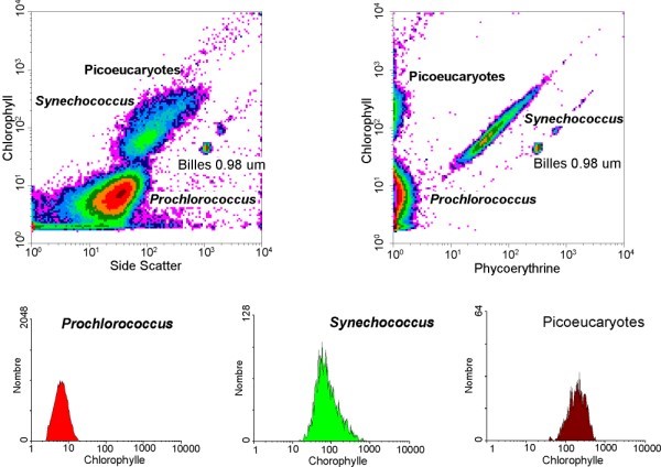
The data is to be analysed by data generated by computer either in histogram or dot plot. Computer analysis give automated population identification, this automated identification could potentially help finding of rare hidden population.
Fluorescence-activated cell sorting (FACS) is a specialized type of flow cytometry. It provides a method for sorting a heterogeneous mixture of biological cells into two or more containers, one cell at a time, based upon the specific light scattering and fluorescent characteristics of each cell. It is a useful scientific instrument as it provides fast, objective and quantitative recording of fluorescent signals from individual cells as well as physical separation of cells of particular interest. The technique was expanded by Len Herzenberg, who was responsible for coining the term FACS.
Fluorescence-activated cell sorting (FACS) is a specialized type of flow cytometry. It provides a method for sorting a heterogeneous mixture of biological cells into two or more containers, one cell at a time, based upon the specific light scattering and fluorescent characteristics of each cell. It is a useful scientific instrument as it provides fast, objective and quantitative recording of fluorescent signals from individual cells as well as physical separation of cells of particular interest. The technique was expanded by Len Herzenberg, who was responsible for coining the term FACS.
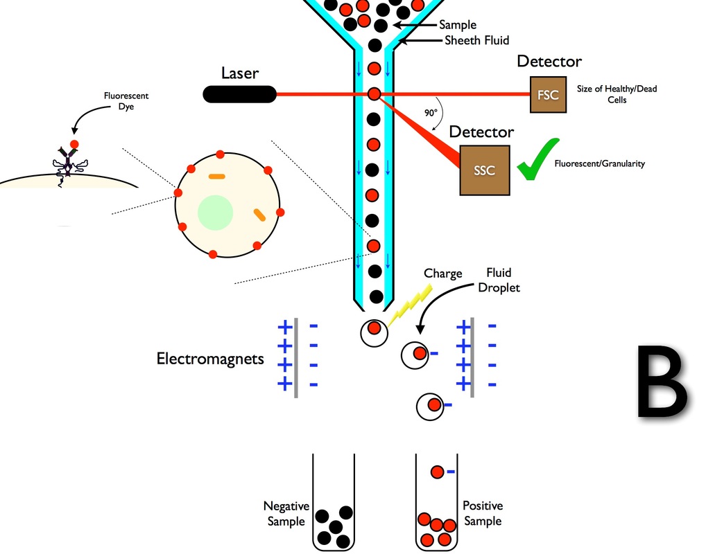
The cell suspension is entrained in the center of a narrow, rapidly flowing stream of liquid. The flow is arranged so that there is a large separation between cells relative to their diameter. A vibrating mechanism causes the stream of cells to break into individual droplets. The system is adjusted so that there is a low probability of more than one cell per droplet. Just before the stream breaks into droplets, the flow passes through a fluorescence measuring station where the fluorescent character of interest of each cell is measured. An electrical charging ring is placed just at the point where the stream breaks into droplets. A charge is placed on the ring based on the immediately prior fluorescence intensity measurement, and the opposite charge is trapped on the droplet as it breaks from the stream. The charged droplets then fall through an electrostatic deflection system that diverts droplets into containers based upon their charge. In some systems, the charge is applied directly to the stream, and the droplet breaking off retains charge of the same sign as the stream. The stream is then returned to neutral after the droplet breaks off
The technology has applications in a number of fields, including molecular biology, pathology, immunology, plant biology and marine biology It has broad application in medicine (especially in transplantation, hematology, tumor immunology and chemotherapy, prenatal diagnosis, genetics and sperm sorting for sex preselection). Also, it is extensively used in research for the detection of DNA damage, caspase cleavage and apoptosis In marine biology, the autofluorescent properties of photosynthetic plankton can be exploited by flow cytometry in order to characterise abundance and community structure. In protein engineering, flow cytometry is used in conjunction with yeast display and bacterial display to identify cell surface-displayed protein variants with desired properties
The technology has applications in a number of fields, including molecular biology, pathology, immunology, plant biology and marine biology It has broad application in medicine (especially in transplantation, hematology, tumor immunology and chemotherapy, prenatal diagnosis, genetics and sperm sorting for sex preselection). Also, it is extensively used in research for the detection of DNA damage, caspase cleavage and apoptosis In marine biology, the autofluorescent properties of photosynthetic plankton can be exploited by flow cytometry in order to characterise abundance and community structure. In protein engineering, flow cytometry is used in conjunction with yeast display and bacterial display to identify cell surface-displayed protein variants with desired properties
Summary of flow cytometry and FACS
Fluorescence activated cell sorting it is part of Flow cytometry. This technique used for the counting, sorting of cell and protein engineering, Based upon their properties of Bio molecules.
- In that techniques laser is going to monitor cell i.e. cells either have high cell weight or light weight.
- Cells containing sample is passed from laser light through niddle like column so as to perfectly get forward scatter value and side scatter value of laser light pattern, These FSC and SSC Detector gathering the data of laser light intensity and it will show the value on computer as graph i.e. graph is plotted FSC Vs SSC and in that at the bottom side strong electromagnet is present this working as a cell sorter
- Electromagnet is going to separate positively and negatively charged cell
- On the basis of graph value of FSC and SSC we can estimate how much amount of cell are healthy and how much are apoptic i.e. the cells are going to shrink and due to that cell shows low FSC and SSC value on the graph.
- It is majorly used in cancer cell, and animal cell culture
- It also contain analogue to digital conversion system which converts fluorescence light in to electrical signal for computer analysis.
- It is also applicable in immunology experiments and plant pathology i.e. disease
- In FACS size of healthy and dead cell is monitored by FSC light and Granularity of cell is monitored by SSC light. It shows the value by histogram and dot plot.
- Cells contains various organelle if healthy cells are present in higher concentration it will indicated by FSC value.


